Products
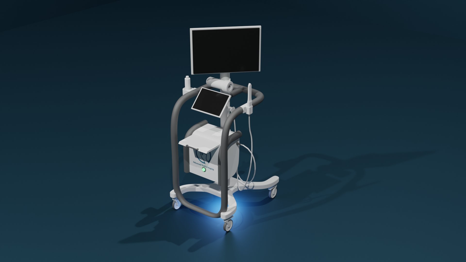
Vaginal Tactile Imager (VTI)
A clinically validated platform to aid in diagnosis and evaluation of vaginal and pelvic floor conditions.
It provides Biomechanical Integrity score (BI-score) as well as quantitative data for:
Vaginal tissue elasticity
Pelvic floor support
Pelvic muscle contraction
Muscle relaxation
Muscle mobility
CPT Code: 0487T
Biomechanical mapping, transvaginal, with report
FDA Approval (USA)
CE Mark (Europe)
TGA Approval (Australia)
Advancements for
Urogynecology
Gynecology
Physiotherapy
Cosmetic Gynecology
Developed with financial support of the National Institute on Aging (NIA) of the National Institutes of Health, USA.

Principle of Operation
The VTI allows acquisition of pressure response patterns during tissue/structure deformations from the vaginal walls along the entire vagina. This enables visualization of vaginal and pelvic floor support structures, and recording of pelvic floor muscle contraction patterns. The VTI software provides data visualization, analysis, information, and reporting tools. The acquired data can then be used for quantitative biomechanical assessment of vaginal and pelvic floor conditions. Among patients, 54% classified the VTI comfort level as more comfortable than manual palpation, 36% as the same, and 10% as less comfortable than manual palpation.
Indications for Use (FDA)
"The Vaginal Tactile Imager obtains a high-resolution mapping of pressures and assesses the strength of pelvic floor muscles within the vagina. It is used in a medical setting to acquire the pressures and store the corresponding data. It also provides visualization, analysis tools and information. The real-time data, as well the analysis information, can then be viewed with an intention of assisting in the diagnosis and evaluation. The device is intended for use by physicians, surgeons and medically trained personnel."
Clinical Utility
The female pelvic structures have varying viscoelastic characteristics across physiological states, and healthy and diseased conditions. The VTI provides a large body of measurements to evaluate individual variations in support defects, as well as identify specific potential markers to measure soft tissue properties and muscle function in patients.
The VTI may be used for:
POP (Pelvic Organ Prolapse): To characterize its development in biomechanical terms.
UI (Urinary Incontinence): To quantify pelvic muscle defects that contribute to UI.
Atrophy: To characterize vaginal tissue atrophy.
Surgery: To provide metrics for pelvic floor surgery outcomes.
Physiotherapy: To monitor pelvic conditions during the course of applied treatment.
RF (Radiofrequency) and Laser treatments: To evaluate vaginal conditions before and after laser or RF treatment.
Military and Sport: To monitor pelvic floor changes under heavy, repetitive loads.
Vaginal Tactile Ultrasound Imager (TIUSv)
A unique device to aid in diagnosis and evaluation of vaginal and pelvic floor conditions. It provides tactile (stress) and ultrasound image (anatomical, strain) fusion using the vaginal probe.
Indications for Use:
“The Vaginal Tactile Ultrasound Imager obtains a high-resolution mapping of pressures, ultrasound images and assesses the strength of pelvic floor muscles within the vagina. It is used in a medical setting to acquire the pressures, ultrasound images and store the corresponding data. It also provides visualization, analysis tools and information. The real time data as well as the analysis information can then be viewed with an intention of assisting in the diagnosis and evaluation. The device is intended for use by physicians, surgeons and medically trained personnel.”
Regulatory status: FDA approved
Developed with financial support of Eunice Kennedy Shriver National Institute of Child Health & Human Development (NICHD) of the National Institutes of Health, USA.