

Technology
Biomechanical Mapping of the Female Pelvic Floor
Definitions
Tactile Imaging
is a medical imaging modality translating the sense of touch into a digital image. The tactile image is a function of P(x, y, z), where P is the pressure on soft tissue surface under applied deformation and x, y, z are coordinates where pressure P was measured. The tactile image is a pressure map on which the direction of tissue deformation must be specified [van Raalte and Egorov, 2015].
Functional Tactile Imaging
translates muscle activity into dynamic pressure pattern P(x, y, t) for an area of interest, where t is time and x, y are coordinates where pressure P was measured.
It may include [Egorov, et. al 2016]:
Muscle voluntary contraction
Involuntary reflex contraction
Involuntary relaxation
Specific maneuvers
Biomechanical Mapping
Biomechanical Imaging = Tactile Imaging + Functional Imaging [Egorov, 2023]
Applications of Biomechanical Mapping (BM) in various pelvic disease conditions:
Prolapse and incontinence: Objective, quantitative pelvic floor assessment — removing guesswork from prolapse and incontinence diagnosis and surgical planning.
Preterm delivery and maternal birth trauma: BM can help predict the risk of preterm delivery and maternal birth trauma by evaluating the biomechanical properties of the cervix, the uterine wall, and the pelvic floor muscles. This information can be used to tailor preventive measures and manage high-risk pregnancies more effectively.
Endometriosis, adenomyosis, and uterine fibroids: BM can help visualize and quantify the extent of these pathologies by assessing the biomechanical properties of the affected tissues, leading to improved diagnosis and treatment planning.
Figure 1. This technology was advanced in cooperation with Artann Laboratories within the scope of the NIA/NIH grants (Phases I-IV).
Figure 2. BI-score and its components for a 58 y.o. patient acquired with the VTI [Egorov, et. al 2022].
Biomechanical Integrity Score (BI-score)
Recently, we have introduced a novel quantitative integral parameter called the Biomechanical Integrity Score (BI-score) to assess and characterize the female pelvic floor. This innovative metric enables the objective evaluation of alterations in pelvic tissues, support structures, and functionalities under various pathological conditions. By utilizing the BI-score, researchers and clinicians can gain deeper insights into the biomechanical transformations of the pelvic floor, facilitating improved diagnosis and treatment of related disorders.
BI-score and What It Reflects
The BI-score is a system measure of the pelvic floor’s ability to bear and distribute functional loads (stress–strain, stress–function mapping).
Low BI-score = weak, overstretched, or damaged support structures (high laxity, poor load transfer, not effective muscle functions).
Normal BI-score = intact fascia–muscle–ligament support system, balanced force distribution, good tissue elasticity, healthy muscles.
Figure 3. A concept of the Digital Twin of the female pelvic floor.
Digital Twin of the Female Pelvic Floor
Digital twin technology, originally developed for intricate physical systems, holds great potential in women’s healthcare, particularly in the management of pelvic floor disorders. It can amalgamate various data sources such as imaging, biomechanical assessments, and patient-reported outcomes to offer personalized diagnostic and therapeutic insights. Through the utilization of 3D modeling and machine learning, the digital twin may facilitate precise visualization, prediction, and individualized treatment planning. It is crucial to address the ethical and practical challenges related to data privacy and ensuring fair access. As this technology progresses, it has the potential to revolutionize gynecological and obstetric care by enhancing diagnostics, customizing treatments, and increasing patient involvement [Egorov, 2024].
Patents
Issued
Method and probe for predicting spontaneous preterm delivery. US Patent No. 12,672,853; July, 2026.
Tactile ultrasound method and probe for predicting spontaneous preterm birth. US Patent No. 12,622,679, May, 2026.
Tactile ultrasound method and probe for predicting preterm birth. US Patent No. 12,564,372; March, 2026.
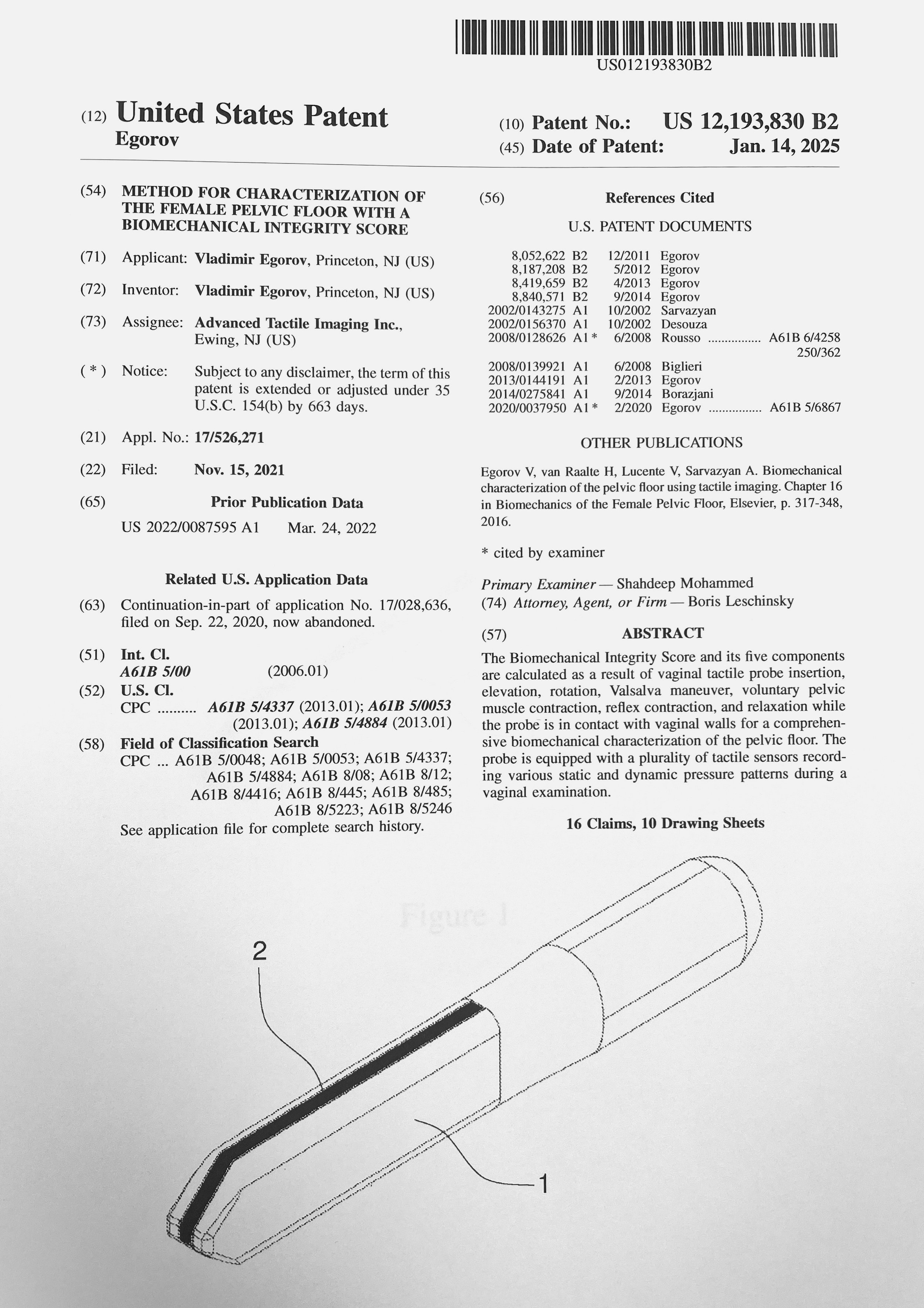
Method for characterization of the female pelvic floor with a biomechanical integrity score. US Patent No. 12,193,830; January, 2025.
Methods and probes for vaginal tactile and electromyographic imaging and location-guided female pelvic floor therapy. US Patent No. 9,861,316; January, 2018.
Method and device for measuring tactile profile of vagina. US Patent No. 8,840,571; February, 2014.
Methods for assessment of improvements in pelvic organ conditions after an interventional procedure. US Patent No. 8,419,659; November, 2013.
Methods for assessment of pelvic organ conditions affecting the vagina. US Patent No. 8,187,208; May, 2012.
Method of characterization and differentiation of tissue. US Patent No. 8,142,368; March, 2012.
Tactile sensor array for soft tissue elasticity imaging. US Patent No. 8,069,735; December, 2011.
Methods for characterizing vaginal tissue elasticity. US Patent No. 8,052,622; November, 2011.
Pending
Digital twin of the female pelvic floor. US Patent Application No. 18,374,721; September 2023.
Tactile ultrasound method and probe for predicting spontaneous preterm birth. US Patent Application No. 18,370,439; September 2023.
Learn More About
Tactile Imaging in Cancer Detection: Sarvazyan, et. al 2011 (review)
Elastography - Emerging Branch of Medical Imaging: Sarvazayn, et al 2011 (review)
Tactile Imaging for Female Pelvic Floor: Egorov, et. al 2016 (book chapter)
Biomechancial Mapping of the Female Pelvic Floor: Egorov, 2023 (book)